1.3 Membrane structure
Essential idea:
The structure of biological membranes makes them fluid and dynamic.
Nature of science:
Using models as representations of the real world—there are alternative models of membrane structure. (1.11)
Falsification of theories with one theory being superseded by another—evidence falsified the Davson-Danielli model. (1.9)
Understandings:
1.3.U1 Phospholipids form bilayers in water due to the amphipathic properties of phospholipid molecules.
Guidance: Amphipathic phospholipids have hydrophilic and hydrophobic properties.
1.3.U2 Membrane proteins are diverse in terms of structure, position in the membrane and function.
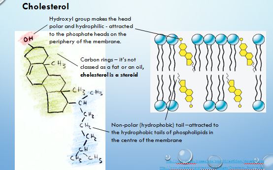
1.3.U3 Cholesterol is a component of animal cell membranes.
Applications & Skills:
1.3.A1 Cholesterol in mammalian membranes reduces membrane fluidity and permeability to some solutes.
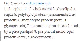
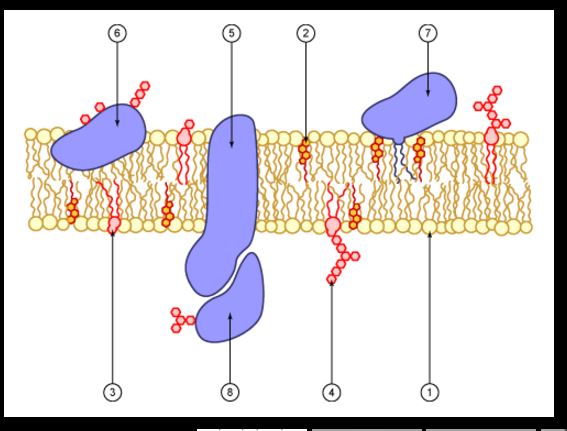
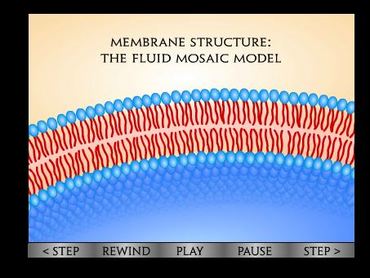
1.3.S1 Drawing of the fluid mosaic model.
Guidance: Drawings of the fluid mosaic model of membrane structure can be two dimensional rather than three dimensional.
Individual phospholipid molecules should be shown using the symbol of a circle with two parallel lines attached.
A range of membrane proteins should be shown including glycoproteins.
1.3.S2 Analysis of evidence from electron microscopy that led to the proposal of the Davson-Danielli model.
1.3.S3 Analysis of the falsification of the Davson-Danielli model that led to the Singer-Nicolson model.
Theory of knowledge:
The explanation of the structure of the plasma membrane has changed over the years as new evidence and ways of analysis have come to light. Under what circumstances is it important to learn about theories that were later discredited?
The structure of biological membranes makes them fluid and dynamic.
Nature of science:
Using models as representations of the real world—there are alternative models of membrane structure. (1.11)
Falsification of theories with one theory being superseded by another—evidence falsified the Davson-Danielli model. (1.9)
Understandings:
1.3.U1 Phospholipids form bilayers in water due to the amphipathic properties of phospholipid molecules.
Guidance: Amphipathic phospholipids have hydrophilic and hydrophobic properties.
1.3.U2 Membrane proteins are diverse in terms of structure, position in the membrane and function.
1.3.U3 Cholesterol is a component of animal cell membranes.
Applications & Skills:
1.3.A1 Cholesterol in mammalian membranes reduces membrane fluidity and permeability to some solutes.
1.3.S1 Drawing of the fluid mosaic model.
Guidance: Drawings of the fluid mosaic model of membrane structure can be two dimensional rather than three dimensional.
Individual phospholipid molecules should be shown using the symbol of a circle with two parallel lines attached.
A range of membrane proteins should be shown including glycoproteins.
1.3.S2 Analysis of evidence from electron microscopy that led to the proposal of the Davson-Danielli model.
1.3.S3 Analysis of the falsification of the Davson-Danielli model that led to the Singer-Nicolson model.
Theory of knowledge:
The explanation of the structure of the plasma membrane has changed over the years as new evidence and ways of analysis have come to light. Under what circumstances is it important to learn about theories that were later discredited?
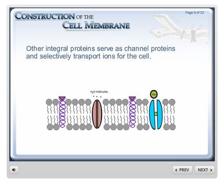
Wisc-Online's Construction of the Cell Membrane is helpful to learn the basics about the structure of the plasma membrane.
|
|
|
A brief look at the fluid mosaic model of the plasma membrane:
|
|
|
1.3.S1 Draw the fluid mosaic model.
Guidance:
|
|
Learn More!
University of Utah Genetics has a page about how membranes help to organize and compartmentalize the cell.
http://learn.genetics.utah.edu/content/cells/membranes/
http://learn.genetics.utah.edu/content/cells/membranes/
